A New Jersey dermatology practice has begun using a device called Nevisense to improve the detection of melanoma, the most dangerous form of skin cancer. Metropolitan Dermatology announced the introduction of the technology, which uses a method called electrical impedance spectroscopy to analyze suspicious moles and lesions on the skin.
According to a report from PR Newswire, Nevisense works by measuring how skin tissue responds to a small, safe electrical current. Cancerous and pre-cancerous cells have different electrical properties than healthy tissue, and the device uses those differences to generate a score that helps dermatologists decide whether a lesion needs a biopsy. The process is non-invasive and takes place during a standard office visit.
Melanoma is responsible for the majority of skin cancer deaths in the United States, even though it accounts for a relatively small percentage of all skin cancer diagnoses. Early detection is the single most important factor in survival rates. When melanoma is caught before it spreads beyond the outer layer of skin, the five-year survival rate is very high. When it is found after it has spread to distant organs, that rate drops sharply.
Dermatologists currently rely on visual examination and dermoscopy, a technique that uses magnification and lighting to examine the structure of a lesion, as the primary tools for evaluating suspicious spots. Those methods depend heavily on clinical experience and can miss lesions that do not have obvious visual warning signs. Nevisense is designed to add an objective, measurable data point to that evaluation process.
The device has been cleared by the U.S. Food and Drug Administration. Clinical studies cited in support of the technology found that it has a high sensitivity for detecting malignant melanoma, meaning it is effective at identifying lesions that turn out to be cancerous. The tradeoff is that it also produces some false positives, flagging benign lesions as potentially concerning, which can lead to biopsies that turn out to be unnecessary.
Metropolitan Dermatology indicated that Nevisense will be used alongside existing diagnostic methods rather than replacing them. The practice presented the technology as an additional layer of analysis that could improve outcomes for patients whose lesions fall into a gray zone where visual examination alone does not provide a clear answer.
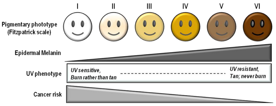
Skin cancer is the most commonly diagnosed cancer in the United States. Dermatologists recommend annual full-body skin checks, particularly for patients with a history of sun exposure, a large number of moles, a personal or family history of skin cancer, or fair skin.