A research team at the University of Hong Kong has developed a new 3D microscopy technique that captures images up to eight times faster than conventional methods, while also exposing biological tissue to significantly less light in the process.
The system, called AIMED, short for Arbitrary Illumination Microscopy with Encoded Depth, was developed in the OMEGA laboratory under Professor Kenneth K. Y. Wong of HKU's Department of Electrical and Computer Engineering. According to a report by Phys.org, the study was published in the journal Advanced Photonics.
The technique targets a well-established problem in biological imaging. Multiphoton microscopy, or MPM, is widely used for three-dimensional imaging deep inside living tissue. It plays an important role in studying neuronal structures, blood vessel networks, and real-time biological activity inside living organisms. But acquiring a full 3D volume with conventional MPM is slow and requires sustained light exposure, which can damage tissue and limits how long researchers can observe a living sample.
Standard MPM works by scanning one thin layer of tissue at a time, stacking those images to build a 3D picture. AIMED abandons that approach entirely. Instead of scanning plane by plane, the system uses a spatial light modulator to split a single laser beam into multiple focal points at different depths, illuminating several layers of tissue simultaneously in one exposure.
The key to separating those overlapping signals lies in the physics of multiphoton excitation itself. Two-photon and three-photon excitation are nonlinear processes that naturally confine their effects to the precise focal point, which reduces interference between the different depth layers being recorded at the same time. The signals from each layer can then be separated computationally using sparse optimization algorithms derived from compressive sensing theory.
Because AIMED captures multiple depth layers in each exposure rather than one, it needs far fewer total exposures to build a complete 3D image. That directly cuts the cumulative light dose delivered to the tissue, which matters for long-term imaging studies where repeated scans over hours or days can otherwise cause photodamage.
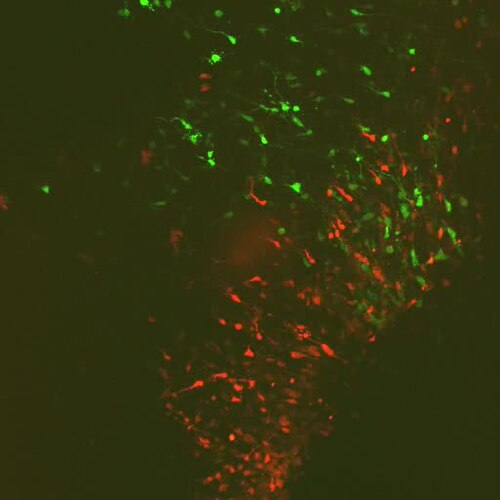
The team validated the system by imaging mouse brain tissue. In a five-plane configuration, the lateral resolution held at approximately 600 nanometers. The researchers also confirmed that intensity across the multiple focal planes remained consistent, with the system able to compensate for the fact that light naturally weakens as it travels deeper into tissue.
The AIMED system was designed to work with existing microscopy hardware without requiring major modifications, which the researchers say makes it more practical for laboratories to adopt. The combination of speed, reduced light exposure, and compatibility with standard equipment positions it as a candidate for studying fast biological processes that current MPM systems cannot capture reliably.