Scientists may have found what has been a missing piece in understanding Alzheimer's disease. Researchers have identified a previously overlooked mechanism of brain cell death that appears to play a major role in both Alzheimer's and frontotemporal dementia, a related condition.
The discovery, reported by Science Daily and based on work from King's College London, centers on a process called karyoptosis. The researchers worked with the UK Dementia Research Institute and received support in part from Alzheimer's Research UK. Their findings were published in Nature Communications.
For decades, scientists have known that neurodegenerative diseases including Alzheimer's, frontotemporal dementia, and ALS involve the buildup of toxic proteins inside neurons. Over time, those nerve cells die, leading to memory loss and other symptoms. Several forms of cell death have been identified over the years, but none fully explained the scale of neuron loss seen in these diseases.
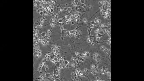
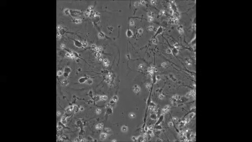
Karyoptosis may be that explanation. The process describes a chain of chemical reactions that begins when toxic proteins accumulate inside a cell. As those reactions unfold, the cell's nucleus, which holds its genetic material, gradually shrivels and eventually breaks apart.
The study analyzed 3,000 brain cells collected from 28 people who had either frontotemporal dementia or end-stage Alzheimer's disease. Using computational algorithms, the research team identified different forms of cell death occurring within the tissue. They found signs of karyoptosis in 35 percent of cells from the frontal cortex of people with Alzheimer's disease, compared with just 15 percent of cells from healthy older adults.
"This study is the culmination of a 10-year journey at King's, from when we first identified karyoptosis in a relatively rare disease to discovering that it is a common feature of dementias which affect millions of people."
Beyond identifying the mechanism itself, the researchers also uncovered a molecular pathway that appears to control it. They found that forcing proteins inside neurons to clump together, which is a hallmark of many neurodegenerative diseases, can trigger karyoptosis. The buildup of toxic proteins destabilizes the outer membrane of the nucleus, causing it to shrink and eventually disintegrate.
The team then turned their attention to proteins called kinases, which function as molecular switches within this pathway. Identifying those switches could give drug developers a specific target for treatments aimed at interrupting karyoptosis before cells are destroyed. No treatments targeting this mechanism currently exist, and the findings are expected to guide future research into slowing the progression of Alzheimer's and related conditions.