

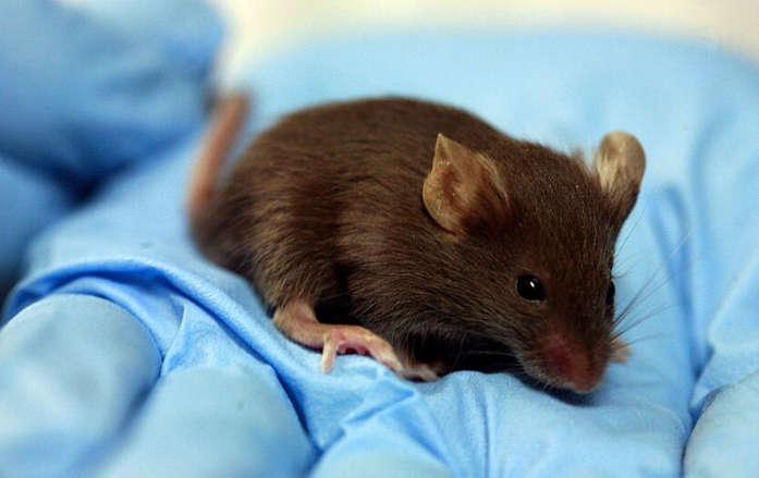
Scientists at Stanford University have developed a new type of mouse that contains significant amounts of human brain tissue. These mice were genetically modified to lack most of their cortex and hippocampus, creating space for human cells to grow. The researchers transplanted human brain organoids into the mice shortly after birth. These organoids were made from skin cells taken from donors and reprogrammed to become brain tissue. Within a few months, the human cells had grown to fill most of the vacant cortical space, taking up about half of the total brain volume. The transplanted tissue connected with the mouse’s blood supply and began to mature. Although the human brain cells were not fully developed, they showed signs of early-stage maturation, similar to what occurs halfway through human pregnancy. The researchers call the animals xenocortical mice. Senior author Sergiu Pașca said descriptions such as mice with human brains are not accurate because the animals still retain a mouse nervous system.
The mice with human brain tissue showed partial improvement on some memory-related deficits compared with mice lacking their cortex and hippocampus, according to the Nature paper. However, the mice lacking their own cortex and hippocampus still acted normally in most ways. They walked around and made sounds, but they had trouble with memory tasks. The results suggest the human grafts can contribute to brain function, but the study did not show that the mice developed human-like thinking. Three to six months after transplantation, the xenocortical mice generally performed similarly to normal mice on behavioral tests.
The researchers believe these mice could help study brain injuries and disorders such as schizophrenia, epilepsy, cerebral palsy, profound autism and some forms of dementia. Pașca's team also noted that the human cells formed rare types of neurons not seen in typical mouse brains, according to a report from The Guardian. These include von Economo neurons, which are also found in humans and other large-brained social animals including great apes, elephants, dolphins and whales. The cells are of particular interest because they are vulnerable in some forms of frontotemporal dementia.
The work was published in the journal Nature. Experts say this technique could change how scientists study the human brain. It allows researchers to grow human brain tissue in living animals and observe its behavior over time. Some scientists have already begun using similar methods to connect brain organoids with computers. Researchers are developing systems that connect organoids to electronic interfaces and use their electrical activity for computing experiments.
The researchers also tested the model by exposing some xenocortical mice to five hours of low oxygen. The human cortical tissue showed substantial injury while ordinary mice were largely unaffected by the same exposure. The xenocortical mice also developed problems with gait and coordination after the low-oxygen exposure. Researchers said the experiment could help scientists study brain injuries associated with oxygen deprivation around birth.
But the approach raises ethical concerns about consciousness and animal welfare. Pașca and Stanford bioethicist Hank Greely helped organize a 2025 meeting with scientists, ethicists and patient advocates to discuss these issues. He said he does not believe the mice have any human-like thinking abilities yet. However, he warned against doing similar experiments on primates.
He considers that a clear ethical boundary for such research. Pașca said transplanting human organoids into monkeys would not be justified at this point. The Nature paper also said future experiments involving more mature grafts or nonhuman primates would require early ethical guidance.
Other scientists say brain organoids grown in dishes could reduce the need for animal experiments, while this new model is intended for questions that require studying human tissue inside a living animal. Still, they emphasize that such experiments must be carefully reviewed and justified. Cambridge researcher Madeline Lancaster told The Guardian that this type of animal experiment needs a strong scientific reason and that many research questions do not require it.
This new model gives researchers a powerful tool to understand how human brain disorders develop.