

Scientists have developed a new method for finding microscopic particles released by brain cells in human blood, potentially opening a new window into diseases that are difficult to detect and study.
The discovery, published October 23, 2026, in Nature Neuroscience, could eventually help researchers investigate Alzheimer's disease, Parkinson's disease and other neurological disorders through ordinary blood samples.
The research team, led by Dmitry Ter-Ovanesyan and colleagues at Harvard-affiliated institutions, identified a protein called NRXN3 on tiny particles known as extracellular vesicles.
These particles are released by cells throughout the body and can carry proteins, RNA and other molecular material.
Researchers demonstrated that they could isolate NRXN3-bearing vesicles from human blood plasma and cerebrospinal fluid. The results establish an important laboratory technique, although further work is needed to prove that the isolated particles originate specifically from neurons.
The study does not introduce a new diagnostic test for brain disease. Instead, it provides researchers with a possible way to examine molecular information associated with brain cells without directly sampling brain tissue.
Tiny Packages May Carry Information From the Brain
Every day, cells throughout the human body release enormous numbers of microscopic particles into surrounding fluids.
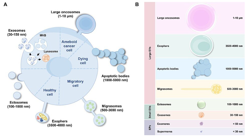
Many of these particles are called extracellular vesicles, or EVs.
They are enclosed by a thin membrane similar to the outer covering of a cell. Inside, they may carry proteins, fragments of genetic material and other molecules associated with the cells that produced them.
Scientists sometimes describe these vesicles as packages that cells send into their environment.
Some participate in communication between cells, while others may help remove unwanted material or influence biological processes.
Because their contents can reflect conditions within the cells that released them, researchers have become increasingly interested in using vesicles as biological markers.
A blood sample contains extracellular vesicles from many different tissues and organs.
The problem is determining where each particle originated.
For scientists studying neurological diseases, identifying particles released by neurons would be especially valuable.
Neurons are the specialized nerve cells responsible for transmitting information throughout the brain and nervous system.
Changes in their proteins, genetic activity and other molecular processes may provide evidence of disease long before serious symptoms develop.
Why Studying the Living Brain Is Difficult
Unlike many other organs, the brain cannot be routinely sampled for laboratory testing.
Doctors can examine blood, urine and other bodily fluids with relatively little difficulty. However, collecting actual brain tissue requires invasive procedures that are inappropriate for most diagnostic purposes.
Cerebrospinal fluid offers another source of information about the nervous system.
This clear fluid surrounds the brain and spinal cord and contains molecules associated with neurological activity.
Collecting it generally requires a lumbar puncture, commonly called a spinal tap.
Although the procedure is widely used in medicine, a blood test would be more convenient for many patients.
Researchers have therefore spent years searching for reliable evidence of brain activity and disease circulating in the bloodstream.
Extracellular vesicles are attractive candidates because some particles and their molecular contents may provide information about processes occurring within the nervous system.
However, scientists must first reliably separate potentially brain-derived material from the enormous amount of unrelated material circulating in blood.
That is the problem addressed by the new research.
The Discovery Centers on a Protein Called NRXN3
The research team investigated proteins that might help distinguish extracellular vesicles associated with neurons from particles released by other cells.
Their approach combined information about gene activity with measurements of proteins found in extracellular vesicles.
The scientists examined large datasets describing which genes are active in different human tissues and cell types.
They then searched for proteins that were particularly associated with neurons and could appear on the surfaces of extracellular vesicles.
Surface proteins are useful because researchers can develop antibodies that recognize and attach to them.
Once an antibody binds to a particle, laboratory equipment can help separate that particle from surrounding material.
The team identified several promising candidates before concentrating on three proteins: SLC12A5, SLC32A1 and NRXN3.
Using sensitive laboratory measurements, the researchers found that NRXN3 was associated with extracellular vesicles in both cerebrospinal fluid and blood plasma.
The other two candidates did not provide equally convincing evidence across the tested human fluids.
This made NRXN3 the leading candidate for further experiments.
How Scientists Separated the Particles
The research involved several laboratory technologies designed to identify and isolate extremely small biological structures.
First, the investigators developed a method for purifying extracellular vesicles from blood plasma and cerebrospinal fluid.
Blood contains large amounts of proteins and other particles that can interfere with measurements.
Removing those contaminants is important because researchers need to know whether a protein is actually attached to a vesicle or simply floating freely in the sample.
The team used a purification approach involving specialized laboratory resin to improve the quality of its samples.
Under certain experimental conditions, the method increased vesicle purity by approximately 100 times.
Researchers then used highly sensitive protein measurements and imaging techniques to examine potential markers on the purified particles.
They developed antibody-based isolation procedures designed to capture vesicles displaying NRXN3.
These procedures successfully recovered NRXN3-positive particles from human plasma and cerebrospinal fluid.
The scientists also examined samples from 18 individuals during part of their imaging validation work.
Together, the experiments demonstrated that NRXN3-bearing vesicles could be detected and separated from complex human biological fluids.
However, detecting NRXN3 does not automatically prove that every captured vesicle came from a neuron.
The researchers emphasized that additional testing of the particles' molecular contents will be needed to establish their cellular origins more firmly.
An Earlier Testing Problem Makes This Discovery Important
The new research addresses a significant problem in previous attempts to identify neuron-derived extracellular vesicles.
Earlier studies frequently used a protein called L1CAM to capture particles thought to originate from neurons.
However, research involving some of the same scientists showed that much of the L1CAM detected in blood plasma and cerebrospinal fluid was present as a free-floating protein rather than attached to extracellular vesicles.
That raised questions about whether certain earlier experiments had isolated the intended particles.
Without reliable evidence of a particle's origin, scientists risk measuring biological material from unrelated sources.
The new study introduced a systematic process for selecting and checking candidate markers.
Rather than relying on a protein's association with neurons alone, the investigators also tested whether it was actually present on extracellular vesicles.
This additional verification could improve the reliability of future research.
Possible Applications in Alzheimer's and Parkinson's Disease
One major potential application involves Alzheimer's disease.
Alzheimer's develops through complicated biological changes involving abnormal proteins, damaged connections between neurons and progressive loss of brain function.
Researchers are investigating blood-based biomarkers that may help identify these processes and measure how they change over time.
If neuronal extracellular vesicles can be isolated reliably, scientists might examine the proteins and RNA they carry for additional evidence of disease.
Similar opportunities exist for Parkinson's disease.
Parkinson's involves the progressive loss of dopamine-producing neurons and is frequently associated with abnormal accumulation of a protein called alpha-synuclein.
Detecting molecular changes associated with affected neurons could help scientists investigate disease development and treatment responses.
Other possible applications include multiple sclerosis, certain psychiatric conditions and neurological injuries.
The new technique has not been shown to diagnose any of these conditions.
Its immediate value is as a research tool that may make it easier to study disease-related molecules in future experiments.
Other Recent Research Supports the Approach
The discovery adds to a growing body of evidence that blood contains molecular information related to brain disorders.
In June 2026, researchers at the Icahn School of Medicine at Mount Sinai published related findings in Nature Communications.
That study examined extracellular vesicles and even smaller particles isolated from human brain tissue and blood.
Researchers identified RNA patterns that distinguished samples associated with Alzheimer's disease from control samples.
Interestingly, some of the smaller particles performed better than conventional extracellular vesicles in certain comparisons.
The findings suggested that information relevant to Alzheimer's disease may circulate in several different types of microscopic particles.
A separate study published in Brain on October 14, 2026, investigated how abnormal alpha-synuclein proteins are associated with extracellular vesicles.
The researchers identified differences among Parkinson's disease, dementia with Lewy bodies and multiple system atrophy.
Some molecular patterns could also be detected in blood, suggesting a possible approach to distinguishing related neurological disorders.
Both studies remain experimental and require additional clinical validation.
Together with the new Nature Neuroscience research, they demonstrate growing scientific interest in examining microscopic particles as sources of information about the nervous system.
Important Questions Remain
Despite the technical progress, researchers still face several obstacles before the method can be used in routine medicine.
One problem involves the NRXN3 protein itself.
Different forms of the protein may exist, including versions that are not attached to extracellular vesicles.
Antibodies used to capture particles could potentially recognize those soluble forms as well.
Another challenge is proving that the recovered vesicles originated from neurons rather than other cells expressing similar proteins.
The researchers also need to determine how consistently NRXN3 appears in samples from people of different ages and health conditions.
The amount and composition of particles circulating in blood may change during illness.
Changes in the blood-brain barrier could also influence how many potentially brain-derived particles reach the bloodstream.
Finally, scientists must establish whether the molecular material captured using the new technique provides reliable information about disease.
Finding a particle in blood is only the first step. Demonstrating that its contents accurately reflect specific changes inside the brain is considerably more difficult.
What Comes Next
The study involved researchers from the Wyss Institute for Biologically Inspired Engineering, Harvard Medical School, the Massachusetts Institute of Technology, the Broad Institute and Brigham and Women's Hospital.
The team developed a computational process that could also help identify extracellular vesicles associated with other brain cells.
These include astrocytes, which support neurons, and oligodendrocytes, which produce the insulating myelin surrounding nerve fibers.
Microglia, the brain's resident immune cells, are another possible target.
Identifying particles from these different cell types could help researchers investigate inflammation, nerve damage and changes in brain function.
Future studies will need to confirm the origin and molecular contents of isolated vesicles and test whether they provide useful information in people with neurological diseases.
The investigators have made their computational approach available to other scientists, providing a starting point for additional research.
For now, the discovery represents an advance in laboratory technology rather than an immediate change in medical care.
Its importance lies in creating a potential way to collect information associated with living brain cells from a blood sample, an approach that could make neurological research less invasive and more accessible.