

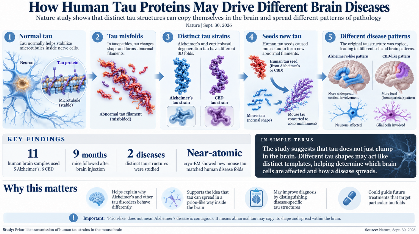
Scientists have shown that abnormal tau proteins taken from human brains can preserve their disease-specific shapes after triggering new tau deposits in mouse brains. The finding provides some of the strongest evidence yet that tau can behave in a prion-like way inside the brain. It may help explain why different tau disorders damage different cells and produce different patterns of disease.
| Study Summary: Researchers found that abnormal tau proteins from people with Alzheimer’s disease and corticobasal degeneration can act like distinct prion-like strains. When these human tau proteins were introduced into mouse brains, they caused the mice’s own tau to form new abnormal structures that closely matched the original human disease. The findings suggest that the shape of tau itself may help determine how different neurodegenerative diseases develop and which brain cells are affected. |
The study was published September 30 in Nature. Researchers used tau taken from people who had Alzheimer’s disease or corticobasal degeneration, a rarer neurological disorder known as CBD.
Tau is normally an important protein inside nerve cells. It helps stabilize microscopic structures called microtubules, which act like internal tracks for moving materials through neurons. In several neurodegenerative diseases, tau changes shape, separates from microtubules, and builds up into abnormal filaments.
These diseases are collectively called tauopathies.
Alzheimer’s disease is the most common tauopathy, but abnormal tau also appears in corticobasal degeneration, progressive supranuclear palsy, Pick’s disease, chronic traumatic encephalopathy, and some forms of frontotemporal dementia. Scientists have known for years that tau does not form exactly the same structure in every one of these diseases.
That difference may matter more than previously understood.
Using cryo-electron microscopy, researchers have found that tau filaments from different diseases fold into distinct three-dimensional structures. A major question has been whether those structures merely accompany different diseases or whether the shape of the tau itself helps determine what kind of disease develops.
The new work directly tested that idea.
Researchers prepared insoluble tau filaments from the frontal cortex of five people with confirmed Alzheimer’s disease and six people with corticobasal degeneration. They then injected the material into the striatum of normal wild-type mice.
The scientists followed the animals for months to see whether the human tau would cause the mice’s own tau protein to form abnormal deposits. It did.
Nine months after injection, both groups of mice had developed abnormal phosphorylated mouse tau in several parts of the brain. The pathology had spread beyond the original injection site into areas that included the cerebral cortex and corpus callosum.
But the pattern was different depending on which human disease supplied the original tau.
Mice given Alzheimer’s tau developed deposits mainly in neurons and their extensions. Mice given CBD tau developed abnormal tau in both neurons and glial cells, including patterns resembling the coiled bodies and astrocytic plaques seen in human corticobasal degeneration.
That suggested that the original tau structure was influencing which cells became affected.
The researchers then addressed an important alternative explanation. The abnormal material found months later might simply have been the human tau that had originally been injected.
Tests showed that this was not the case. The injected human tau could be detected immediately after injection, but it was no longer detectable after about one week. New abnormal tau deposits appeared later and consisted of the mice’s own tau protein. This means the human tau acted as a seed. It appears to have recruited normal mouse tau and caused it to assemble into abnormal filaments.
The most important part of the experiment came when researchers examined those newly formed mouse filaments at near-atomic resolution with cryo-electron microscopy.
Mouse tau triggered by Alzheimer’s disease seeds adopted the Alzheimer-type fold. Most of those filaments were paired helical filaments, the characteristic structure associated with Alzheimer’s disease.
Mouse tau triggered by CBD seeds instead formed structures matching the tau fold found in human corticobasal degeneration.
The similarities were extremely close at the atomic level.
This is what makes the findings important to the prion-like model of neurodegeneration. A true strain-like process requires more than a protein simply causing another protein to clump. The structural information of the original seed must also be copied into newly formed protein assemblies. The researchers found that this happened with tau.
A prion is a misfolded protein capable of forcing normally folded versions of the same protein into the abnormal shape. Different prion shapes can act as different strains, producing different patterns of damage.
The new study suggests that tau can use a closely related molecular process.
That does not mean Alzheimer’s disease is contagious.
Classical prion diseases such as Creutzfeldt-Jakob disease can, under unusual circumstances, be transmitted between individuals. There is no evidence that ordinary Alzheimer’s disease, corticobasal degeneration, or other common tauopathies spread from person to person through normal contact.
The term “prion-like” describes the way abnormal proteins can seed and reproduce their shapes within the nervous system.
According to the National Institute on Aging, healthy tau normally helps stabilize microtubules inside neurons. In Alzheimer’s disease, abnormal chemical changes cause tau to detach and join with other tau molecules, eventually contributing to neurofibrillary tangles.
Scientists have also observed that tau pathology tends to spread through the brain in recognizable patterns as neurodegenerative disease progresses.
The new findings offer a possible molecular explanation for part of that pattern.
Different tau folds may have different abilities to enter cells, recruit normal tau, reproduce themselves, leave a cell, and move into connected cells. They may also interact differently with neurons, astrocytes, oligodendrocytes, and other cells in the brain.
Those differences could help determine where a tauopathy develops and what symptoms eventually appear.
The study also strengthens the usefulness of certain mouse models. The newly produced mouse filaments closely reproduced the structures of human Alzheimer’s and CBD tau, giving researchers a way to investigate disease-specific tau behavior in a living brain.
There are limits to the model.
Adult mice naturally produce only the four-repeat form of tau, known as 4R tau. Humans produce both three-repeat and four-repeat forms. That means ordinary mice cannot reproduce every human tauopathy equally well.
Pick’s disease, for example, mainly involves 3R tau. Alzheimer’s disease contains both 3R and 4R tau, while corticobasal degeneration primarily involves 4R tau.
The mice in this study also did not develop the severe neurological illness seen in people with advanced tauopathies. They accumulated abnormal tau, but appeared healthy during the study period.
Researchers said this could mean tau propagation and tau toxicity are partly separate processes. It is also possible that the animals did not live long enough, or did not accumulate enough abnormal tau, to develop major symptoms.
The study does not show exactly why one tau structure prefers certain cells or why one disease progresses faster than another. Those are now major questions.
Scientists want to understand which receptors allow tau seeds into cells, what conditions allow a seed to convert normal tau, and how newly formed filaments leave one cell and reach another.
If different tau strains truly help determine different diseases, recognizing those strains may eventually improve diagnosis. It could also affect treatment strategies, because a therapy that blocks one tau structure may not work equally well against another.
The work therefore moves the field beyond the idea that tau accumulation is simply a common feature of several brain diseases. It suggests that the shape of abnormal tau itself can carry biological information. That shape can be copied, preserved, and expressed as a distinct pattern of pathology in another brain.
For researchers trying to understand Alzheimer’s disease and related disorders, that provides a clearer molecular explanation for how one protein can be involved in several very different diseases.