

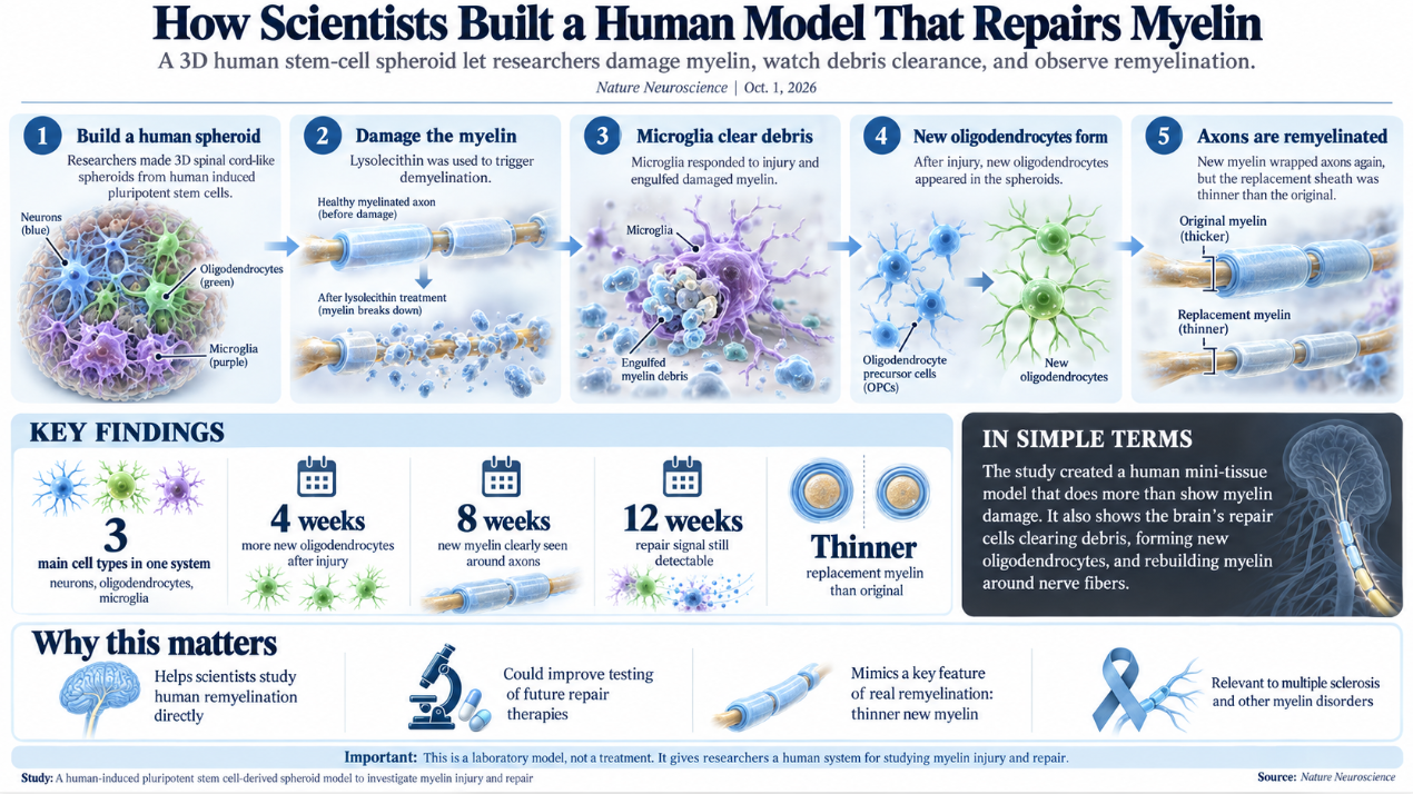
Scientists have built a three-dimensional human cell model that can reproduce myelin damage and then repair it, giving researchers a new way to study a process that is central to multiple sclerosis and other neurological diseases. The model contains human neurons, myelin-producing oligodendrocytes, and functioning microglia. After researchers damaged its myelin, newly formed oligodendrocytes wrapped axons in replacement myelin.
The study was published October 1 in Nature Neuroscience. The researchers used induced pluripotent stem cells, or iPSCs, which are adult cells that have been reprogrammed into a stem-cell-like state and can then be directed to develop into different cell types.
Myelin is a fatty, insulating material wrapped around many nerve fibers in the brain and spinal cord. It allows electrical signals to move efficiently along axons, the long extensions that neurons use to communicate.
Oligodendrocytes make this myelin in the central nervous system.
When myelin is damaged, nerve signaling can slow or fail. Continued myelin loss can also leave axons vulnerable to permanent damage.
This is a central problem in multiple sclerosis, or MS. In MS, the immune system attacks structures in the central nervous system, including myelin. According to the National Institutes of Health, nearly 1 million people in the United States and about 3.1 million people worldwide are affected by MS.
The human nervous system does have some ability to repair myelin. This process is called remyelination. However, the repair often becomes incomplete or less effective as disease progresses.
Studying remyelination in people has been difficult because researchers cannot directly observe all of the cellular events taking place inside living brain and spinal cord tissue. Much of what scientists know about the process has therefore come from animal models.
The new model was designed to provide a human alternative.
The researchers created spinal cord-like spheroids from human iPSCs. These small, three-dimensional structures contained neurons and large numbers of mature oligodendrocytes capable of producing myelin.
They then added cells that developed into microglia.
Microglia are immune cells that live in the central nervous system. They respond to injury, remove damaged material, and can influence both inflammation and tissue repair.
The presence of active microglia made the model more complex than a simple culture containing only neurons and oligodendrocytes. It allowed researchers to follow several parts of the damage-and-repair process in the same human-derived system.
The team damaged the myelin using lysolecithin, also called LPC. This chemical is commonly used in laboratory studies to cause demyelination.
The treatment caused extensive fragmentation of myelin inside the spheroids.
The microglia responded to the injury and began engulfing myelin debris. This is an important part of repair because damaged myelin must be cleared before new myelin can effectively form.
The scientists then watched what happened over the following weeks.
Four weeks after the injury, the spheroids contained significantly more newly generated oligodendrocytes than untreated controls. By eight weeks, many of those new cells had started producing myelin around axons.
The effect remained detectable at 12 weeks.
Nature Neuroscience reported that the newly formed myelin met several structural requirements expected of genuine myelin. The sheaths were connected to oligodendrocytes, formed organized segments around axons, and contained compact layers of membrane.
Electron microscopy allowed the researchers to examine the repaired axons in much greater detail.
The replacement myelin was real, but it was not identical to the original coating.
The repaired axons had thinner myelin sheaths than comparable axons that had not been damaged.
That finding is important because thin myelin is a known feature of remyelinated tissue. It indicates that the system is not simply producing more myelin proteins. It is reproducing a recognizable form of biological repair.
The study also showed that some molecular changes inside the spheroids resembled changes previously observed in human MS tissue.
After the injury, researchers used single-cell RNA sequencing to examine how individual cells changed their gene activity. Oligodendrocytes, precursor cells, and microglia showed shifts in genes and pathways associated with myelin damage and repair.
Among the changes were alterations involving genes connected to oligodendrocyte structure and remyelination.
The researchers also tested the model with clemastine, a drug that has previously attracted attention in remyelination research. In the spheroids, clemastine increased the proportion of newly generated oligodendrocytes that went on to produce myelin.
The experiment was not intended to establish clemastine as a treatment. Instead, it showed that the spheroid system can respond to a substance already known to influence oligodendrocyte biology.
That makes the model potentially useful for screening future compounds.
PubMed describes the system as a platform for studying otherwise inaccessible parts of human myelin biology and for investigating disease mechanisms relevant to multiple sclerosis.
The need for better human models has been a persistent problem in this field.
Animal experiments have identified many biological pathways that appear to stimulate myelin repair. But results in animals do not always translate successfully to people because human cells can behave differently.
A laboratory system built entirely from human-derived cells could allow researchers to test potential repair strategies earlier and more directly.
The model also allows different cell types to be observed together. That matters because remyelination is not controlled by oligodendrocytes alone.
Microglia must remove debris. Oligodendrocyte precursor cells must multiply and mature. New oligodendrocytes must then locate exposed axons and build functional myelin around them.
The researchers were able to watch several of those stages occur in sequence.
There are still important limits.
A spheroid is not a complete human brain or spinal cord. It does not reproduce blood flow, the full immune system, the enormous variety of cells found in living tissue, or the long-term course of multiple sclerosis.
The scientists also used a chemical to create myelin damage. MS involves a much more complicated immune process.
Even so, the system provides something researchers have lacked: a reproducible human model in which myelin can be damaged, cleared, rebuilt, and then examined at the cellular and ultrastructural levels.
The Florey Institute, where many of the researchers are based, has been developing stem-cell and three-dimensional culture models to study myelin and myelin-producing cells in neurological disease.
The new work takes that effort a step further by showing a complete repair response after a controlled injury.
For people with MS, the research does not represent a new treatment.
Its immediate value is as a research tool.
Current MS therapies can reduce inflammation and disease activity, but there is still a major need for approaches that restore lost myelin and protect vulnerable nerve fibers.
By giving scientists a human system in which they can watch remyelination happen, measure where it succeeds, and see where it falls short, the model could make that search more precise.
The thinner replacement myelin may be especially useful. Researchers can now ask why repaired sheaths remain thinner, whether that matters for long-term nerve function, and what might make the repair more complete.
Those questions were previously much harder to study directly in human cells.
The result is a laboratory model that does more than imitate myelin damage. It also shows the nervous system trying to repair itself.